News and Articles
Stay informed with the latest insights from Clemex, a leader in advanced image analysis and microscopy solutions. Our blog features company news, industry trends, technical expertise, and real-world case studies demonstrating how image analysis technology helps laboratories and industrial organizations achieve precise, reliable results across a wide range of applications.
2024-10-21
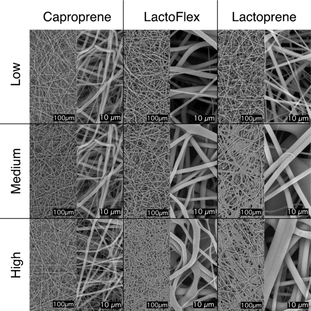
Poly-Med’s Breakthrough in Medical-Grade Scaffolds
Discover how Poly-Med partnered with Clemex Technologies to revolutionize tissue engineering via machine learning and electrospun scaffolds.

2024-03-24
Image analysis using Clemex Studio's artificial intelligence
Recent advances in artificial intelligence have given the possibility for image analysis to be simplified. Learn how by reading this white paper.

2022-09-30
Image analysis for microscopy made simple with machine learning
Automate your QA lab without the complexity. See how machine learning simplifies phase extraction and workflow automation for metallurgists.

2020-09-29
Image Analysis Systems from Clemex - manual vs. automated
Manual microscopy in quality control (QC) saps resources. Learn how automated image analysis systems streamline workflows and maintain accuracy.

2020-09-10
Design-Based Stereology and Binary Image Histomorphometry in Nerve Assessment
Comparing design-based stereology with semi-automated binary image histomorphometry for quantifying healthy peripheral nerve characteristics.

2020-06-04
Calibrating Your Microscopy Systems: How & Why?
Why and how to calibrate your microscopy system with a fully traceable stage micrometer to keep measurements accurate, repeatable, and standards-compliant.

2019-06-11
Automated analysis of laser grooves on wafers
How Clemex Vision automates microscope inspection of laser grooves on silicon wafers, improving die-separation quality and measurement accuracy.

2018-11-21
Automated sulfides quantification by multispectral optical microscopy
A study on identifying and quantifying six common sulfide minerals on a polished section using multispectral optical microscopy and automated image analysis.